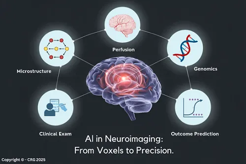
Artificial intelligence is transforming neuroimaging from a static interpretive tool into a dynamic, integrated component of neurologic care. By combining voxel-level analytics with clinical, physiologic, and biomarker data, AI enables a modern anatomoclinical approach in which imaging functions as bedside neuropathology. Near-term advances in quantum sensing are expanding the range of measurable phenomena, while longer-term developments in quantum computing promise to enhance modelling and prediction. Together, these developments position neuroimaging as both a clinical and strategic asset for modern health systems.
Artificial intelligence (AI) has rapidly become the driving force behind the modernisation of medical imaging, and nowhere is this more evident than in neuroimaging. Once limited to static visual interpretation, imaging now serves as a computational foundation for diagnosis, triage, forecasting, and system-level decision support. Early AI tools demonstrated that algorithms could detect hemorrhages, infarcts, or tumors with remarkable speed and accuracy. Today, the field stands at the threshold of something far more expansive: the integration of imaging with clinical, genetic, physiologic, and operational data into unified, predictive models of neurologic disease.
For hospitals and health systems, this shift marks the transition from imaging as a diagnostic endpoint to imaging as a strategic asset, one that accelerates care, optimises resource utilisation, and strengthens institutional performance.
The first wave of AI-powered imaging tools focused on recognition: identifying critical abnormalities quickly and reliably. That chapter is now well established. Platforms equipped with deep learning detect large-arterial occlusions, flag intracranial hemorrhages, gauge aneurysm growth over time, and quantify tumor burden with sensitivity that often enhances human performance. These tools have reduced preliminary read times, improved after-hours coverage, and served as an indispensable safety net in high-volume clinical environments.
But the true transformation lies in what modern platforms do beyond detection. AI-enabled systems extract quantitative features from every voxel, subtle textural, structural, metabolic, and microvascular patterns that elude human perception. Machine-learning models then map these features against clinical trajectories, linking imaging signatures to outcomes, recurrence risk, or treatment response.
The result is not simply a “faster read,” but a deeper read, one that converts visual data into prognostic intelligence.
For practicing clinicians, especially neurologists who routinely interpret their patients’ imaging studies, AI-driven neuroimaging has become an inherent component of the clinical encounter itself. Historically, imaging lived adjacent to bedside assessment, functioning as a confirmatory or localisation tool. Today, AI dissolves that separation. Modern multimodal platforms integrate structural imaging, perfusion metrics, physiologic signals, biomarker profiles, and clinical narratives into a consolidated framework aligned with the clinician’s real-time understanding of the patient.
This convergence represents a contemporary evolution of Charcot’s Anatomoclinical Method. Where Charcot correlated observable signs with pathologic anatomy at autopsy, clinicians now correlate bedside findings with computationally derived imaging biomarkers, effectively practicing Bedside Neuropathology. AI strengthens this correlation by extracting microstructural and microvascular patterns that reflect underlying disease biology, enabling neurologists to map clinical syndromes directly to their pathophysiologic substrates.
As a result, imaging is no longer a static artifact; it becomes a living component of clinical reasoning. AI contextualises the picture with the patient’s clinical trajectory, allowing clinicians to integrate anatomy, physiology, and probability into a single diagnostic and prognostic narrative. For hospital leaders, this represents more than a technological upgrade; it signals a shift toward an integrated model of neurologic care in which imaging is a continuous partner in clinical decision-making.
In acute stroke care, where every second has neurologic consequences, AI-enabled neuroimaging has become indispensable not only for systems-level efficiency but for the vascular neurologist’s bedside decision-making. Automated perfusion and arterial analysis platforms provide rapid, standardised characterisation of the ischemic core, penumbra, early infarction, and vessel status. Yet their greatest value lies in their pairing with the vascular neurologist’s intimate understanding of the patient’s examination, comorbidities, and evolving clinical picture.
This integration modernises the Anatomoclinical Method approach. The vascular neurologist correlates subtle motor findings, gaze or language deficits, and visual field defects with AI-derived perfusion maps and tissue viability scores. What once required inference and probability becomes a precise synthesis: imaging biomarkers align with physiologic observations to guide eligibility for thrombolysis, thrombectomy decisions, and recovery projections.
Neuropathology. It reveals tissue viability, vascular dynamics, and metabolic compromise at moments when they matter most. This convergence strengthens rapid triage, expands access to expert-level stroke interpretation, and improves accuracy across diverse teams, while preserving the vascular neurologist’s central role as interpreter of disease trajectories.
For executives, AI-enabled neuroimaging is not solely a clinical asset; it is a strategic operational lever that shapes workforce design, quality assurance, patient safety, and digital transformation.
AI serves as a force multiplier in high-volume imaging environments. Automated detection, triage, and quantification systems automate repetitive analytical tasks that traditionally fall to clinicians. This redistributes cognitive labor, reduces burnout, and permits neurologists and imaging specialists to focus on complex interpretive challenges and patient-facing decisions. In distributed or rural health systems, AI standardises interpretation across campuses, ensuring tertiary-level analytic support everywhere.
Hospitals are increasingly measured on standardisation, documentation accuracy, and adherence to evidence-based pathways. AI reduces inter-reader variability, produces reproducible quantitative measurements, and embeds structured reporting aligned with accreditation, regulatory expectations, and value-based reimbursement models. Automated volumetrics, perfusion thresholds, and lesion detection drive internally consistent quality dashboards and audit processes.
Diagnostic error, particularly missed or delayed findings, remains a significant source of liability. AI provides a parallel safety layer, flagging critical abnormalities, alerting clinicians to discrepancies, and supporting consistent triage during off-hours. Integrated into enterprise risk-management strategies, AI reduces medico-legal exposure and signals proactive adoption of technologies that mitigate preventable harm.
The full value of AI emerges when neuroimaging analytics permeate the health system’s digital ecosystem. When linked to electronic medical records (EMRs), transfer centers, teleconsultation workflows, scheduling platforms, stroke-alert systems, and command centers, AI ensures imaging insights trigger operational action, mobilize teams, accelerate transfers, and coordinate bed or operating room (OR) scheduling. Imaging becomes a core component of enterprise IT strategy.
Institutions that view imaging not as a standalone clinical service but as a foundational digital infrastructure will hold a distinct advantage as computational intelligence becomes a defining feature of system performance.
As AI grows more sophisticated, neuroimaging faces two parallel frontiers:
Quantum sensing represents the most immediate and transformative advance in neuroimaging. Unlike quantum computing, which remains limited by hardware constraints, quantum sensors are rapidly approaching clinical feasibility. Technologies such as optically pumped magnetometers (OPM-MEG), nitrogen-vacancy (NV) diamond magnetometry, and hybrid quantum–optical sensors are demonstrating profound capabilities:
These capabilities are not speculative; they exist now in research and preclinical domains and are poised to enter advanced clinical workflows within the coming decade. Quantum sensing therefore, redefines neuroimaging’s near-term trajectory by enhancing the resolution, portability, and physiologic fidelity of what can be measured at the bedside.
Quantum computing remains early in its developmental arc but holds long-term promise for accelerating complex modeling tasks central to neuroimaging and neurologic disease simulation.
It is essential to note that fully real-time, whole-brain network simulations are not yet feasible. No existing quantum computer has the qubit count, noise tolerance, or algorithmic framework for such modeling. However, emerging hybrid quantum–classical approaches offer targeted accelerations in specific computational domains:
As quantum hardware matures, improving qubit coherence, error correction, and circuit depth, these targeted accelerations could evolve into more adaptive and higher-fidelity models of brain network behavior.
Together, quantum sensing and quantum computing delineate a dual-path future:
One reshaping measurement, the other reshaping modeling.
Neuroimaging is evolving from illustration to computation, from images to insight. AI does not replace the neuroimaging specialist; it amplifies expertise, accelerates decision-making, and broadens institutional capability across both clinical and administrative domains.
For clinicians, it delivers precision.
For executives, it offers a strategy.
For patients, it ensures faster, safer, and more individualised care.
The story of AI in neuroimaging is ultimately the story of medicine becoming more integrated, anticipatory, and intelligent, transforming voxels into precision and information into impact.
NOTE: This is the second in a series of four articles that comprehensively cover the different dimensions and the forecasted outcome of AI in medical practice.

Dr. Qureshi is one of the most senior interventional neurologists in the United States and a widely published researcher. In addition to being a Professor at the University of Missouri, he is also the Director of the Zeenat Qureshi Stroke Institutes and the Editor-in-Chief of the Journal of Neuroimaging. He has been a collaborator in various projects studying AI in clinical medicine.

Dr. Geyer is a seasoned neurologist with subspecialty expertise in epilepsy and sleep disorders. Currently, he is a Professor in the University of Alabama College of Community Health Sciences and Director of the Advanced Imaging Facility and MRI Research Facility for the University. He has a unique interest in the application of AI to all aspects of clinical medicine.

Dr. Gong is an Associate Professor of Computer Science at The University of Alabama and the founding Director of the Alabama Center for the Advancement of Artificial Intelligence (ALAAI). A recognised leader in AI research, education, and innovation ecosystems, he has spearheaded initiatives that link advanced AI methods with pressing societal challenges, including rural healthcare resilience, environmental monitoring, and entrepreneurship education.

Paul Cox is a healthcare technology leader with deep expertise in AI/ML, bioinformatics, and software development. He has directed the research and commercialization of advanced AI models that drive data-driven healthcare innovations across medical devices, diagnostics, digital health, and microbiome therapeutics. Currently, he is CEO of Evimero and leads the bioinformatics efforts to develop next-generation tools for disease diagnostic devices, precision therapeutics, and human health optimization.
Dr. Gomez, a pioneer in vascular, critical care, and interventional neurology, has spent over four decades advancing stroke treatment and mentoring physicians. A passionate educator, he challenges conventional thinking through engaging lectures and his protean publications. As a Professor of Neurology at the University of Missouri, he continues to advance the novel applications of AI in clinical medicine.