

In recent years, a number of targeted and immuno-oncology drugs have been developed and used to treat lung cancer patients, resulting in improved outcomes for lung cancer patients, but cancer mortality from lung cancer has not improved. What is the underlying reason for this? The majority of innovative cancer drugs are prescribed for patients with stage IV metastatic NSCLC. While they have significantly increased survival from an average of 1.5 years to 3-4 years, the vast majority of patients with metastatic lung cancer do not achieve a cure, relapse due to resistance, and usually die. Even if some patients are cured, the quality of life of surviving advanced lung cancer patients is very poor due to the various side effects and complications that occur during treatment.
So what is the best way to reduce the mortality rate of lung cancer, especially non-small cell lung cancer, which accounts for the majority of lung cancer patients? The answer is self-evident. It is early lung cancer diagnosis. The five-year survival rate for patients with NSCLC is directly proportional to the stage. Stage I is 70-90%, Stage II is 60%, Stage III is 30-40%, and Stage IV is 0-10%. Therefore, if the number of patients diagnosed at stage I increases, the number of patients with stage IV metastatic lung cancer will naturally decrease, and lung cancer mortality will decrease. However, the current stage distribution of NSCLC patients at diagnosis is approximately 25-30% stage I, 5-10% stage II, 20-25% stage III, and 40-50% stage IV, which means that the majority of patients are diagnosed at stage III or higher, and the stage I diagnosis rate, which has a high cure rate, is low. Therefore, increasing the stage I diagnosis rate is a key strategy to reduce lung cancer mortality.
So why is the diagnosis rate for early stage lung cancer so low? Since the National Lung Screening Trial (NLST) study in 2011, which reported a 20% reduction in lung cancer mortality in high-risk individuals, such as smokers, using low-dose computed tomography (LDCT), early lung cancer screening using LDCT has become commonplace, but the false positive rate is so high that it is difficult to determine whether a lung lesion detected by LDCT is lung cancer or not. In order to confirm lung cancer, pathological findings by histological or cytological examination of the suspected lesion are essential. Lung biopsy is therefore essential. Lung biopsy is not difficult for large tumours in stage 2 and above, but stage 1 lung cancer is often small in size and dangerous in location. Ground-glass, cystic, or consolidation type nodules, which are common in Asians, are difficult to obtain adequate tissue and are at high risk of complications. Recently, efforts have been made to diagnose peripheral lung nodules by histological examination using advanced techniques such as navigation bronchoscopy, peripheral bronchoscopy with radial EBUS, and robotic assisted bronchoscopy, but these techniques require general anaesthesia, are expensive, and diagnostic performance is not cost-effective even when performed by experts. In general, for stage I NSCLC, the majority of patients are diagnosed by frozen biopsy in the operating theatre, and if confirmed as lung cancer, lung cancer surgery is usually followed. Therefore, about 15% of patients who undergo surgical biopsy for suspected cancer are found not to have cancer, and the diagnosis of lung cancer by surgical biopsy can lead to delayed diagnosis, which can lead to postoperative recurrence.
Unfortunately, patients who are diagnosed with early stage lung cancer and undergo surgery face the problem of recurrence. Postoperative adjuvant anti-cancer treatment only provides a survival gain of 5.4%, and patients with stage I can only be followed up. Osimertinib (Tagrisso), a targeted anti-cancer drug, has been used for three years as postoperative adjuvant therapy for stage 2 and 3 EGFR gene mutation-positive patients, but this is insufficient and is currently being studied for five years, and there is still no prevention of recurrence for stage 1 NSCLC patients, who make up the majority of surgical patients. Neoadjuvant immunochemotherapy has recently gained attention to reduce recurrence after NSCLC surgery. Patients with resectable NSCLC who do not have driver oncogene such as EGFR or ALK mutation before surgery are treated with a combination of cytotoxic chemotherapy and immune check-point inhibitor before surgery, and then undergo surgery, and the efficacy of neoadjuvant therapy has been demonstrated to prevent postoperative recurrence by shrinking tumours, downstaging, and eliminating micrometastases. The complete pathological response (CPR) and major pathological response (MPR) rates, which are surrogate markers of neoadjuvant therapy, have been shown to be more than 20% and 50%, respectively. In contrast, neoadjuvant therapy with osimertinib in EGFR-mutated NSCLC has not yet become a standard of care due to low MPR rates.
In summary, targeted agents and immunotherapy, which have been prescribed for advanced stage 3 and 4 NSCLC, are being extended to early stage 1 and 2 operable patients, highlighting the importance of histological diagnosis and genetic and molecular testing to identify driver oncogenes in patients with suspected early stage lung cancer. Given the importance of neoadjuvant treatment, it is necessary to confirm the histological diagnosis and genotype for EGFR mutations by biopsy before surgery. Accurate EGFR genotyping is necessary to avoid unnecessary neoadjuvant immunotherapy in patients with EGFR mutations because EGFR-mutated NSCLC is refractory to immunotherapy. In recent years, there have been attempts in the United States to introduce robotically assisted bronchoscopy for pre-operative histological diagnosis and genetic/molecular testing in patients with lung nodules detected on low-dose CT (LDCT). However, it requires expensive equipment and is performed under general anesthesia. There is a great unmet need for innovative diagnostic methods for stage I lung cancer as histological diagnosis and genetic and molecular testing are invasive and sometimes dangerous, and a significant proportion of patients are diagnosed and treated through surgical biopsy. Against this background, there has been a lot of development of liquid biopsy using blood, but the diagnostic sensitivity of plasma liquid biopsy using cfDNA is very low, around 20%.
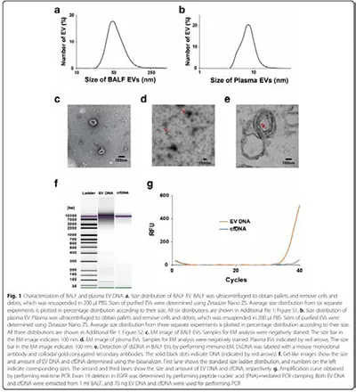
To compensate for this, the author developed an innovative lung cancer diagnostic method called Exosome-based BALF liquid biopsy (ExoBAL). It is an innovative and noble liquid biopsy method that diagnoses lung cancer by separating exosomes or extracellular vesicles (EVs), which are messengers of cancer cells, from BAL Fluid (BALF) obtained by performing bronchoalveolar lavage (BAL) through bronchoscopy near the lesion observed on chest CT and analysing exosomal DNA, RNA, and proteins carried by exosomes. Cancer cells secrete a much larger amount of exosomes than normal cells, and bioactive molecules such as DNA, RNA, and proteins are packaged in exosomes according to the characteristics of the parent cell, so analysing exosomes can identify the characteristics of the cancer cells that secrete them. Since BALF contains exosomes secreted by cells comprising the tumour microenvironment (TME) such as immune cells, epithelial cells, endothelial cells and fibroblasts in addition to tumour cells, it is possible to assess the TME of lung cancer without the need for cancer tissue, making ExoBAL a very good liquid biosource for simultaneous analysis of tumour and TME.
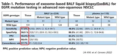
The authors discovered the presence of oncogenic mutant dsDNA in exosomes in laboratory studies using EGFR-mutant lung cancer cells and were the first to identify and report the presence of EGFR-mutant DNA in exosomes from BALF obtained from EGFR-mutant NSCLC patients. Based on these findings, we developed a diagnostic method to test EGFR genotype through ExoBAL instead of invasive lung biopsy to identify cancer cells in lung cancer diagnosis. EGFR mutation testing is an essential genetic test that must be performed in patients diagnosed with lung cancer, and currently, if lung cancer cells are confirmed by histological examination, DNA is extracted from FFPE and performed by PCR method, which requires a turnaround time of more than two weeks, and results cannot be obtained if cancer cells are not confirmed by histological examination. ExoBAL, on the other hand, is fast and accurate, with a sensitivity, specificity and concordance of 95% within 1-2 days in advanced NSCLC patients, and has the advantage of obtaining results without invasive biopsy. In a study comparing the performance of tissue vs. ExoBAL EGFR genotyping in 224 advanced NSCLC patients, only 5 patients had discordant results, showing a sensitivity, specificity, positive and negative predictive rate and concordance rate of more than 97%, and ExoBAL's performance in more than 1000 advanced NSCLC patients over the past 5-6 years has shown a tissue concordance rate of more than 95%, and is currently awaiting approval by the Korean FDA.
Based on the successful performance in these advanced NSCLC patients, we are currently conducting a study to detect stage I EGFR mutant lung cancer without invasive tissue biopsy or surgical biopsy using ExoBAL, which is highly effective in detecting EGFR mutant cases, especially in small sized nodules, ground-glass type, partly solid nodules, cystic or pneumonic type lesions where lung biopsy is not possible or dangerous. In Asian countries, including South Korea, lung cancer in non-smokers and women accounts for more than 35% of all lung cancers, and 60-70% of these are EGFR mutation-positive lung adenocarcinomas. We are conducting a clinical study in which the 3rd generation EGFR-TKI lazertinib is administered as neoadjuvant treatment for 9 weeks, followed by restaging and surgery to confirm pathology and tissue EGFR genotyping in ExoBAL-detected EGFR mutant patients without biopsy, and adjuvant lazertinib is administered for 3 years if pathological staging is confirmed as 2 or higher. The study is entitled Neoadjuvant lazertinib therapy for potentially resectable EGFR-mutated NSCLC detected by ExoBAL (NCT 05469022). We recently announced the interim results of the study, which identified 48 EGFR mutant ExoBAL-detected patients from 128 screened patients, 40 of whom started neoadjuvant lazertinib treatment and 34 of whom underwent surgery. Of the 34 patients who underwent surgery, 33 were diagnosed with adenocarcinoma and one with adenosquamous carcinoma, and EGFR genotyping revealed subtype concordance in all but one patient, for a concordance rate of 97.1%. In addition, down-staging was confirmed in 55% of patients, with a 60-80% down-staging benefit in clinical stage IB and above. 33 patients had complete R0 resection and one patient had R1 resection, who is receiving lazertinib as adjuvant postoperatively. The overall response rate was 66%, which was slightly lower due to the inclusion of stage I patients, and there were several cases of unresectable cases converted to resectable cases in clinical stage III patients. However, the MPR rate was low at 18.4%, similar to the sub-20% MPR rate seen in other neoadjuvant therapies with osimertinib. Although the low MPR rate, which is a surrogate marker of neoadjuvant therapy, has not attracted attention to neoadjuvant therapy with EGFR-TKIs, it can be inferred that increasing the duration of neoadjuvant treatment to 6 months or more, or combining neoadjuvant treatment with chemotherapy or amivantamab, may improve the MPR rate and reduce the postoperative recurrence rate and increase the survival rate of many early-stage EGFR mutant NSCLC patients. In practice, the postoperative recurrence rate of EGFR mutation-positive surgical patients is very high, ranging from 30% in stage I, 4-50% in stage II, and 7-80% in stage III. While adjuvant treatment with osimertinib has become the standard of care for stage 2 and 3 patients, there is no special treatment for stage 1 lung cancer patients, who make up the majority of surgical patients. We believe that it is clinically important to identify stage 1 EGFR mutant cases with a high likelihood of recurrence before surgery, and we expect that if ExoBAL can identify EGFR mutant cases in stage 1 lung cancer patients with difficulty in preoperative histological examination and provide effective neoadjuvant treatment and surgery, it can reduce the postoperative recurrence rate and ultimately reduce mortality.
Following the EGFR mutation test, we are currently developing a test to detect KRAS mutant cases without tissue biopsy using a similar method, and it is expected that ExoBAL will also be able to detect fusion oncogene such as ALK and ROS1. In addition, we are developing an ExoBAL diagnostic method using miRNA profiling for the remaining 50% of NSCLC patients in whom no driver oncogene is detected, and expect to complete an innovative diagnostic platform for early diagnosis and treatment of stage I lung cancer without tissue biopsy within 2-3 years.
References.
1. Hur JY, Kim HJ, Lee JS, Choi CM, Lee JC, Jung MK, Pack CG, Lee KY. Extracellular vesicle-derived DNA for performing EGFR genotyping of NSCLC patients. Mol Cancer. 2018 Jan 27;17(1):15.
2. Hur JY, Lee JS, Kim IA, Kim HJ, Kim WS, Lee KY. Extracellular vesicle-based EGFR genotyping in bronchoalveolar lavage fluid from treatment-naive non-small cell lung cancer patients. Transl Lung Cancer Res. 2019 Dec;8(6):1051-1060
3. Wu YL, Tsuboi M, He J, John T, Grohe C, Majem M, Goldman JW, Laktionov K, Kim SW, Kato T, Vu HV, Lu S, Lee KY, Akewanlop C, Yu CJ, de Marinis F, Bonanno L, Domine M, Shepherd FA, Zeng L, Hodge R, Atasoy A, Rukazenkov Y, Herbst RS; ADAURA Investigators. Osimertinib in Resected EGFR-Mutated Non-Small-Cell Lung Cancer. N Engl J Med. 2020 Oct 29;383(18):1711-1723.
4. Hur JY, Lee KY. Characteristics and Clinical Application of Extracellular Vesicle-Derived DNA. Cancers (Basel). 2021 Jul 29;13(15):3827.
5. Kim IA, Hur JY, Kim HJ, Kim WS, Lee KY. Extracellular Vesicle-Based Bronchoalveolar Lavage Fluid Liquid Biopsy for EGFR Mutation Testing in Advanced Non-Squamous NSCLC. Cancers (Basel). 2022 May 31;14(11):2744.
6. Kim IA, Hur JY, Kim HJ, Kim WS, Lee KY. A prospective phase 2 study of expeditious EGFR genotyping and immediate therapeutic initiation through extracellular vesicles (EV)-based bronchoalveolar lavage fluid (BALF) liquid biopsy in advanced NSCLC patients. Transl Lung Cancer Res. 2023 Jul 31;12(7):1425-1435