
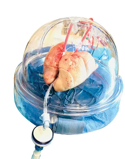
The most effective therapy for patients with end-stage disease is organ transplantation. Lung organ procurement rates from deceased donors for transplantation are significantly lower than those for other solid organs. This low rate is likely due to potential complications that may arise before or after brain death in the donor. These complications include aspiration, thoracic trauma, ventilator-associated barotrauma injury, ventilator-associated pneumonia, and neurogenic pulmonary edema. This results in a shortage of donor lungs leading to a 20% mortality in the lung transplant waiting list. Multiple methods are used to expand the donor pool with the use of extended criteria donors, donation after cardiac death (DCD), aggressive use of Extracorporeal Membrane Oxygenation (ECMO) post-transplantation for marginal lungs, lobar lung transplantations for small thoracic cavity patients, and the use of EVLP. EVLP is a groundbreaking therapy used to treat donor lungs outside the body (ex vivo) prior to transplantation. This process boosts organ quality and transforms previously unsuitable lungs into viable options for transplant.
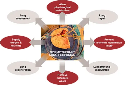
The commonly used method for preserving and transporting lungs historically has been static cold storage (SCS). However, several limitations are associated with SCS. These include tissue damage from prolonged hypothermic preservation, challenges in assessing donor organ function and viability, the effects of ischemia-reperfusion injury (IRI), and limited opportunities for organ repair. EVLP not only serves as a great assessment tool but also enables graft repair, reconditioning, and immunomodulation. EVLP is transforming the landscape of lung transplantation through its ever-expanding applications by improving the transplantability of marginal donor lungs, prolongation of preservation, reconditioning of unsuitable lungs using therapeutics for the treatment of pulmonary emboli, bacterial colonization, pulmonary edema, aspiration injury, and further optimization of suitable lungs like blood group conversion, immunomodulation, virus inactivation, gene therapy, and tissue engineering.
History of Lung Preservation
The original method for preserving isolated organs before SCS began in the 1960s was machine perfusion using plasma or blood-based solutions. Numerous preservation methods were attempted including hyperbaria, hypothermia, and hypothermic pulsatile or non-pulsatile perfusion. Organ preservation was revolutionized with the advent of SCS as scientists hypothesized that lower temperatures could offer cytoprotection by lowering cellular metabolism. It then became the gold standard for preserving organs at hypothermic temperatures. The earliest concepts underwent numerous modifications and refinements, leading to the current practices of organ perfusion and preservation.
In 1935, Carrel and Lindbergh performed the first normothermic ex vivo organ perfusion by explanting the thyroid glands of cats and rabbits and perfusing them for up to a week. In the 1970s, EVLP was tested in various animal models like mice, rabbits, guinea pigs, dogs, and pigs. Hardesty attempted to study normothermic EVLP in the 1980s clinically but had to abandon it due to suboptimal outcomes. Steen et al. revisited EVLP in the mid-1990s to evaluate the transplant suitability of lungs procured from a non-beating heart donor. This trailblazing technique led to the first-in-human lung transplantation from a DCD donor in 2001, following successful evaluation ex vivo. Furthermore, in 2005, the same team went on to perform the first lung transplantation of a marginal lung which was initially rejected and then reconditioned using EVLP. In 2008, Cypel et al. took EVLP a step further and proposed extended EVLP up to 12 hours in a pre-clinical model using a new protocol.
Machine Perfusion
Machine perfusion is an innovative method that optimizes organ condition by circulating a controlled flow of perfusate. This technique significantly enhances organ quality for transplantation purposes. It ensures the delivery of oxygen and nutrients to support tissue metabolism and maintains the tone of the microvasculature in the organs while effectively removing toxic metabolic waste. Different temperatures have been explored for ex vivo machine perfusion. These include normothermic machine perfusion at 35–38 °C, sub-normothermic machine perfusion at 20–34 °C, controlled oxygenated rewarming at 8–20 °C, and hypothermic machine perfusion at 0–8 °C. Normothermic machine perfusion maintains the organ at body temperature under physiologic conditions to preserve metabolic activity and viability.
EVLP Indications
Currently, there are no set standard guidelines for using EVLP. However, EVLP is commonly considered in cases where the PaO2/FiO2 ratio is ≤300 mmHg, there is an expected long period of ischemia, it involves DCD donors, or there are worrisome findings from bronchoscopic evaluation. Poor lung compliance, atelectasis, pulmonary edema, and pulmonary embolism are suitable lung conditions for EVLP. However, mechanical lung injury with air or blood leak, significant contusion, active infectious disease, major aspiration, and malignancy are contra-indicated.
EVLP methodology
There are currently three EVLP protocols used globally- Lund protocol, Toronto protocol, and Organ Care System Lung. The original protocol is the Lund Protocol from Sweden which was further adapted into the currently widely used Toronto protocol. The third protocol involves the use of a portable device that effectively reduces the cold ischemic time. Currently, there are four commercially available EVLP devices in the US that use one of the three standard protocols or a hybrid of them. These devices are Lung Assist™ by Organ Assist®, XVIVO Perfusion System (XPS) ™ by XVIVO® (Figure 02), XVIVO LS™ by XVIVO®, and OCS™ by TransMedics®. In collaboration with Traferox Technologies Inc., the Toronto team successfully engineered the second generation of the TorEx Lung Perfusion System. This remarkable advancement specifically targets and resolves the engineering design limitations that were inherent in the original Toronto EVLP system. (Figure 02)
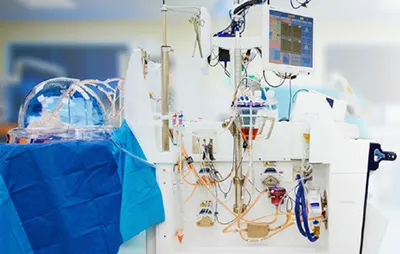
During the process of EVLP, the lungs are kept in a warm (37°C) environment, supplied with a cell-free perfusate solution, and ventilated using a lung-protective ICU-type ventilator. Steen Solution™ is a precisely formulated solution, extensively utilized for EVLP and machine perfusion of various organs. This solution contains colloid components such as human serum albumin and Dextran 40, which help maintain oncotic pressure. Additionally, it includes specific physiological ion concentrations, which collectively play a crucial role in regulating osmolality to maintain the balance. Buffers help to stabilize and uphold normal pH levels, ensuring the organ's internal environment remains within the optimal pH range. Furthermore, the presence of glucose in the solution serves as a vital energy source to support various cellular functions and metabolic processes.
The EVLP system comprises various components in an enclosed and controlled unit to accede to the perfusion and respiratory requirements. The fundamental elements of an EVLP circuit consist of a pump, reservoir, perfusion solution, ventilator, oxygenator, endotracheal tube, air filter, and oxygen sensor. Additionally, the package consists of a container of deoxygenating gas, a set of tubing, and cannulae that need to be connected to both the pulmonary artery and the left atrial cuff.
The surgical part of EVLP consists of suturing the left atrial and pulmonary artery cannula (Figure 03), intubating the trachea, and connecting the cannulae to the lines in the XVIVO circuit after dividing and de-airing the tubings.
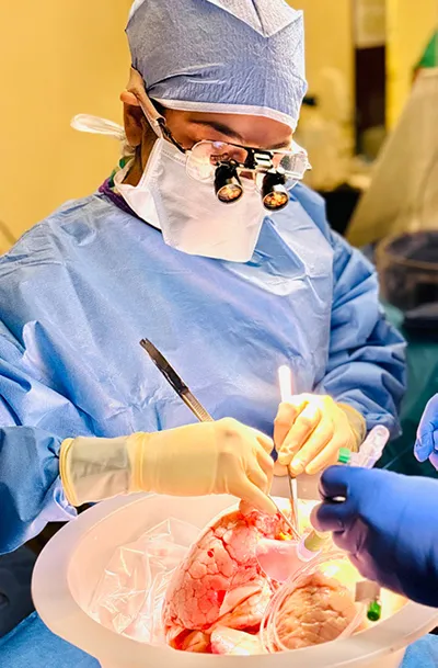
Lung monitoring currently involves assessing physiological parameters such as gas exchange, compliance, and airway pressure; biochemical factors like glucose, lactate levels, pH, and acid-base balance; imaging techniques such as radiographic images and bronchoscopy; and biological measurements including cytokines and chemokines.
The criteria for determining transplant suitability after EVLP usually include stability of lung function parameters (such as pulmonary vascular resistance, lung compliance, airway pressures, and pulmonary artery pressure), improvement in P/F ratio to more than 300 mmHg, and satisfactory lung examination. Conversion rates after EVLP have varied from 34% to 96% across studies. The average overall conversion rate in the United States is 61.8%
Lung repair
Initially, the process of EVLP was limited to a short evaluation period after DCD to determine the suitability of the lungs for transplantation. However, advancements in techniques and technology have made it possible to extend the time for perfusing lungs outside the body, enabling a more thorough and objective assessment of lung quality. This extended evaluation period also allows for interventions to improve the function of the lungs before transplantation.
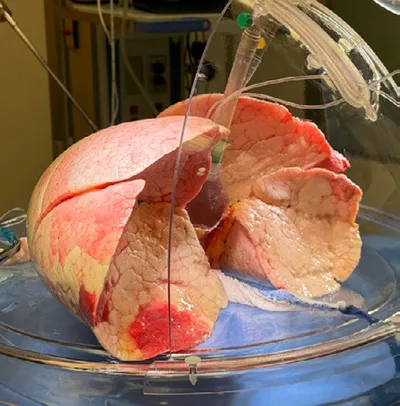
Different drugs have been delivered through the perfusate to prevent IRI. Inhalational gases (NO, CO, H2) have been used during EVLP to reduce inflammatory response and lung edema. Lung injury because of infection and endotoxins has been treated with mesenchymal stem cells. IL-10 gene therapy has made headway to mitigate IRI. When the specific types of injury are identifiable, utilizing injury-specific treatments during EVLP can lead to more effective and targeted care. For example, the addition of high-dose, broad-spectrum anti-microbial agents to the perfusate helps to treat donor lung infection. In one study, acid aspiration-induced lung injury in pigs was effectively treated through lung lavage and surfactant replacement. Furthermore, pulmonary thrombolysis can be performed through EVLP to address pulmonary embolism, followed by a successful lung transplantation. ( Figure 04)
Lung immunomodulation
During EVLP, targeted immunomodulation using RNA-based therapies can be specifically administered to modify the immune response of the donor lungs. This approach aims to reduce the recipient's immune system recognition and sensitization while potentially increasing tolerance markers. As a result, there is potential for a decrease in both acute and chronic rejection, which may lead to a decreased need for long-term immunosuppression.
Outcomes
EVLP has been linked to outstanding short-, mid-, and long-term survival in all studies. Data from retrospective studies conducted at individual institutions and post hoc analyses of clinical trials indicate that there is non-inferior 12-month allograft and patient survival following EVLP when compared to conventional transplantation. Recipients who receive EVLP lungs experience comparable long-term survival without chronic lung allograft dysfunction (CLAD) when compared to recipients who receive conventional lungs.
EVLP and AI
EVLP is distinctly suitable for Artificial Intelligence (AI) and Machine Learning (ML) approaches because the ex vivo data is: (i) limited to an isolated organ and free of mixed signals from other organ systems; (ii) gathered longitudinally over several hours, allowing for a potential trajectory of improvement or deterioration in organ quality, and (iii) derived from numerous monitoring systems, resulting in a high volume of data. Although EVLP offers a potential solution to the organ shortage issue, its effectiveness is hindered by the absence of standardized criteria for determining when an organ is suitable for transplantation. The decision-making regarding EVLP is primarily subjective and involves numerous measurements during the perfusion, which can be overwhelming for inexperienced EVLP programs. To mitigate this, Keshavjee et al. from Toronto developed a machine-learning model in 2023, named InsighTx, to aid surgical decision-making and predict post-transplant outcomes, using clinical EVLP data collected over a decade. A sequential donor-recipient model was developed that could be used for evaluating donor lungs as a general model for any recipient or could be customized with specific recipient details to personalize predictions for individual patients.
Future applications
The potential applications of EVLP are not limited to organ evaluation and the extension of ischemic time; they also encompass the integration of advanced diagnostic and therapeutic modalities. These may include improved transplant logistics (multi-day lung preservation pairing with 10℃ lung preservation with short cycles of EVLP), the opportunity to explore organ banking concepts, better immunological matching between donors and recipients, the opportunity to perform time-dependent therapeutics like blood group conversion, elimination of CMV pre-transplant, and further metabolic rehabilitation.
Cutting-edge research is currently exploring advanced imaging techniques like magnetic resonance imaging and ultrasound to provide a more comprehensive understanding beyond conventional chest radiography during EVLP. The assessment of perfusate samples in preclinical models has conclusively identified the presence of extracellular biomarkers and pro-inflammatory gene expression. Once validated, certain monitoring modalities could play a crucial role in evaluating organ response to ischemia, quantifying inflammation, and predicting future reperfusion injury and/or alloimmune response.
A promising approach to increase the availability of suitable lungs for transplantation involves bioengineering from a scaffold, which serves as a platform for the regeneration of new lungs. The creation of whole-lung extracellular matrix scaffolds involves perfusing cadaveric organs with decellularizing detergents. The EVLP technique can be utilized as a bioreactor with vascular perfusion and liquid ventilation. This helps in repopulating the native lung matrix with endothelial and epithelial cells in the necessary numbers and appropriate anatomic locations, which are essential for enabling organ function. The rat and porcine transplantation models have shown exciting initial progress, demonstrating clear evidence of gas exchange after reperfusion. However, long-term functionality has yet to be attained due to the underdevelopment of the vascular bed and distal lung epithelia. The Toronto research group is currently exploring an innovative method involving the creation of a 3D-printed lung scaffold, known as Ulung. This scaffold has the potential to be infused with either allogeneic cells or a patient's cells. This neoteric technique could pave the way for the creation of personalized and functional lung tissue for transplantation or regenerative medicine applications. It’s crucial to emphasize that much more additional basic and translational research is required before this method can be applied to humans clinically.
Barriers to adoption
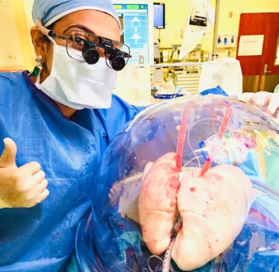
The widespread use of EVLP is limited due to costs. Incorporating EVLP into a lung transplant center's practice requires a significant investment of time and resources. Broadly, the components can be categorized into personnel costs, system costs (equipment and supplies), and physical space. Depending on the chosen EVLP system, there may be a substantial initial capital investment for equipment and hands-on staff training. This requires a strong institutional commitment to the growth of the lung transplant program. It may be challenging for institutions to justify the use of a resource-intensive system that is only used sporadically. For institutions unable to make a large initial investment, outsourcing to a dedicated EVLP facility might be a more practical option. In some cases, a combination approach could offer the most flexibility for a program.
( Figure 05).
Conclusion
The remarkable advancements in Lung preservation research have greatly improved the success of clinical Lung transplantation for patients with end-stage lung disease. However, the high demand for Lungs surpasses the number of available donors. The implementation of EVLP offers a promising strategy to utilize marginal donors by reducing IRI and supporting the repair and regeneration of less optimal grafts. It is also important to optimize current perfusion solutions by adding essential components to meet metabolic needs. Prolonged EVLP creates opportunities for organ repair and reprogramming, highlighting the need for further study of new strategies to enhance the quality of donor grafts before transplantation. This approach contributes substantially to expanding the donor pool, increasing lung transplant volumes, and enhancing overall graft function post-transplantation.