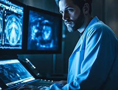
Recent advancements Artificial Intelligence (AI) advancements in machine learning have achieved tasks once reserved exclusively for human ability. Radiology is at the forefront of software tools to improve patient diagnosis and treatment of disease, including in AI. Despite initial fears that these more powerful tools will replace the role the radiologists, AI has become the next generation of tools to support a subspecialty under growing demand for increased medical imaging. AI technology--despite its limitations--may still be at the precipice of greater transformative opportunities, but with radiologists in control to potentiate their positive impact in patient care.
With the rapid development and innovations in machine learning, Artificial Intelligence (AI) has developed several meanings. While the general perception of the definition means a generalized understanding of a problem or process to be able suggest solutions that formerly required human input or cognition. This is more commonly referred to as generalized AI. In practice, AI is about pattern recognition, and exposing or “training” an algorithm or network with a data set to be able to detect these patterns when introduced to novel data.
AI in radiology is no exception. The initial research implementing AI was on nodule detection in mammography to identify areas that represent a higher probability of malignancy. The first FDA approved cloud based deep learning software tool for MR cardiovascular imaging in 2016. This decade is defined by an explosion of AI tools with the promise of exponential improvements in speed and performance with the potential to revolutionize radiology or even eliminate the need for the field entirely. Radiology was not the only specialty at risk of being replaced, as IBM’s AI Watson Health, having proven its language processing skills beating the world’s best jeopardy player in 2011, was showing promise in complex treatment recommendations in oncology to make individualized treatment plans at Memorial Sloan Kettering Cancer Center. Prominent computer scientist Geoffrey Hinton known for his contributions to neural networks once said,
“I think if you work as a radiologist, you are like the Wilie E Coyote in the cartoon. You are already over the edge of the cliff, but you have not looked down yet. There is no ground underneath. People should stop training as radiologists now. It’s just completely obvious that in five years, deep learning is going to do better than radiologists.”
This was said in November of 2016 at a “machine learning and Market for Intelligence seminar.” This warning was so powerful some believe it partially contributed to the reduced training of radiologists for several years. Despite the AI hype, we still have human doctors and radiologists who are increasingly needed in the modern healthcare landscape. However, this does not mean Hinton’s predictions on radiology are principally incorrect, rather premature, and overstated. Transformer machine learning networks can perform better at more tasks than ever before. While the distant future of this technology in radiology is challenging to predict, over the next few years AI will offer exciting opportunities for radiologists to make transformative changes on how modern radiology is performed.
Machine learning tools have shown impressive capabilities in the fields of literature, education, interpretation, art, and data summarization. The initial challenge is the acquisition of training data containing sufficient imaging pathology for new pattern recognition. The data is manually curated to prevent exposure to poor quality data to reduce bias. The most expensive step is training the neural network to refine the prior correlation. Even if all this is done successfully, it must perform at a level that provides utility to the radiologist. Without effortless PACs integration and high sensitivity and specificity, the tools will go unused. The improvement process is not easy because the network nodes are a “black box,” and simple node modulations will not provide the desired effect. Lastly, healthcare policy lags behind these technologies, further slowing down their implementation. The ethical implications and legal repercussions of AI-driven errors limits providers and companies willing to take on the risks with a lack of clear standards and precedents. Radiological AI progress is slower than anticipated, but steadily becoming more prevalent in radiological practice every year. AI now elicits excitement for how the role of the radiologist will transform throughout the years.
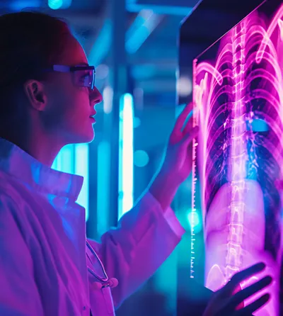
Imaging tools are widely available in every hospital system and are instrumental in numerous treatment and diagnostic evaluation algorithms. Furthermore, the accessibility of volumetric imaging studies has made their implementation more pervasive. As a result, the number of images per case continues to rise every year and increasing volume throughput demands of the available radiologist workforce. AI tools serve an immediate beneficial role as an additional auditor to reduce diagnostic errors and report variability.
The use of AI for interpretive assistance is present today, with the largest proportion of approved algorithms focusing on breast, lung, neurological, and musculoskeletal image interpretation. Breast and lung imaging focus on malignant detection and characterization from various imaging modalities, where neurological algorithms and MSK detect not only concerning masses but also other pathology such as hemorrhage and fractures. Their utility only exists as an aid, as radiologists are on average more sensitive and specific than the AI tool, but offer the most benefit to more junior and trainee radiologists.
Despite impressive computer vision for image interpretation, many are excited to automate the mundane task of protocolling studies. While Watson wasn’t the healthcare moonshot, its pattern detection has the potential to dynamically protocol studies to physician preference. While the correct study is context dependent, the limited number of potential protocol options helps improve the chances of successfully being first line. Furthermore, AI may reduce the burden of documentation. Several institutions implement documentation templates to standardize reporting and encourage a complete search pattern. AI would take this aid a step further by communicating imaging findings automatically into these templates. These are some of the newest use cases being rolled out at academic medical centers due to the rapid advancement of machine learning to perform natural language processing tasks quickly and effectively.
In addition to improving the speed of work, AI prioritization can improve the quality of the radiologist’s work. AI scan prioritization helps the patient and primary care teams by first reading the scans with the most time sensitive findings. Preliminary screening and pattern recognition of ordering context can help reduce the time before communicating critical imaging findings to the primary team. It can also improve acquisition quality and time, with the approved algorithms that can upscale a lower quality pixelated image. When applied to positron emission tomography, shorter detection times reduces child and adult agitation with similar diagnostic capabilities.
The success of these software-based tools is their hyper focused scope. When targeted at niche tasks, they offer an advantage to the radiologist to generate quality image interpretation with improved efficiency. The nascent AI tools still offer significant potential to transform the field and allow the next generation of radiologists to take the reigns toward a bright future in an integral role of the healthcare team.
Similar to radiological images, image interpretation involves many grey areas requiring clinical correlation. Visualization alone can identify over 20,000 pathological states with over 50,000 causal relations to disease. This degree of variability and evolution of imaging technology changes the standards of practice and are in perpetual state of flux. AI must be similarly flexible, plastic, and responsive to increase the breath of its utility. The European Society of Radiology and the American College of Radiology conducted surveys to evaluate how AI tools are being integrated into clinical practice. The overwhelming majority of clinicians reported that these tools did not improve efficiency, performed inconsistently, and failed to be helpful in most cases. The most cited reason for this failure is that the algorithms are brittle, and crack under evolving clinical conditions and software environment. After training these models are static because of the risk of continuous training integration runs the risk of introducing erroneous and incorrect associations.
The AI machine learning networks, and the associated regulations, must evolve its adaptability standards to refine its capability over time in a way that does not introduce unnecessary risk to patients. The primary issue is that this requires significant manual labor to maintain a ground truth state without injecting any bias into an updated training set.
The greatest immediate benefits can be quickly realized in non-interpretive tasks for clinic and procedures. Integrating large language models, such as ChatGPT, can coordinate routine tasks such as patient referral, scheduling, procedural consent, research enrollment, outpatient monitoring, and follow-up. This has a disproportionate impact on procedural focused subspecialties such as Interventional Radiology (IR) but can serve similar functions for radiologists in interdisciplinary teams as a consultant.
While interpretive AI functions exist now, it has not reached the envisioned potential of the synthetic radiologist to interpret and report on new images. For this to be achieved, there must be a concurrent ecosystem of AI algorithms to have access to and compare image interpretation. Their training exists in isolation and will become much stronger if able to competitively refine its interpretation skills against other algorithms and experienced radiologists. Perhaps a public database of old and new radiological images grounded by experienced radiologist interpretation available for AI interpretation and re-evaluation will accelerate the rate at which these networks can read new images as fast and accurately as possible.
AI has captured the imagination of millions to perform tasks once thought impossible without human intervention, and that potential is no greater than in the field of Radiology. The private development of AI tools is fraught with overspeculation, fear mongering, false promises, and failures, but the resurgence of ever more skilled and agile AI software tools continues to fulfil this original promise of revolutionizing the field of radiology.
Current tools provide beneficial widgets to optimize workflow empower the next generation radiologist in an industry acquiring more images per capita than any time in history. The greatest opportunities are yet to come, and envisioning the distant role of this emerging technology is nebulous, the field is on the precipice of the greatest transformative changes to come. Ironically this transformation may actually free radiologists from ever growing tedium and complexity of the current working environment to once again focus on the fundamentals of the subspecialty—applying imaging tools to diagnose and treat disease.
Citations:
1. Driver, C.N., Bowles, B.S., Bartholmai, B.J., and Greenberg‐Worisek, A.J. (2020). Artificial Intelligence in Radiology: A Call for Thoughtful Application. Clin. Transl. Sci. 13, 216–218. https://doi.org/10.1111/cts.12704.
2. Benjamens, S., Dhunnoo, P., and Meskó, B. (2020). The state of artificial intelligence-based FDA-approved medical devices and algorithms: an online database. Npj Digit. Med. 3, 1–8. https://doi.org/10.1038/s41746-020-00324-0.
3. Lohr, S. (2021). What Ever Happened to IBM’s Watson? N. Y. Times.
4. Brady, A.P. (2021). Artificial Intelligence in Radiology: An Exciting Future, but Ethically Complex.
5. Mello-Thoms, C., and Mello, C.A.B. (2023). Clinical applications of artificial intelligence in radiology. Br. J. Radiol. 96, 20221031. https://doi.org/10.1259/bjr.20221031.
6. Vasey, B., Ursprung, S., Beddoe, B., Taylor, E.H., Marlow, N., Bilbro, N., Watkinson, P., and McCulloch, P. (2021). Association of Clinician Diagnostic Performance With Machine Learning-Based Decision Support Systems: A Systematic Review. JAMA Netw. Open 4, e211276. https://doi.org/10.1001/jamanetworkopen.2021.1276.
7. Trivedi, H., Mesterhazy, J., Laguna, B., Vu, T., and Sohn, J.H. (2018). Automatic Determination of the Need for Intravenous Contrast in Musculoskeletal MRI Examinations Using IBM Watson’s Natural Language Processing Algorithm. J. Digit. Imaging 31, 245–251. https://doi.org/10.1007/s10278-017-0021-3.
8. Liao, Y., Liu, H., and Spasić, I. (2023). Deep learning approaches to automatic radiology report generation: A systematic review. Inform. Med. Unlocked 39, 101273. https://doi.org/10.1016/j.imu.2023.101273.
9. Yu, F., Endo, M., Krishnan, R., Pan, I., Tsai, A., Reis, E.P., Fonseca, E.K.U.N., Lee, H.M.H., Abad, Z.S.H., Ng, A.Y., et al. (2023). Evaluating progress in automatic chest X-ray radiology report generation. Patterns 4. https://doi.org/10.1016/j.patter.2023.100802.
10. Chilamkurthy, S., Ghosh, R., Tanamala, S., Biviji, M., Campeau, N.G., Venugopal, V.K., Mahajan, V., Rao, P., and Warier, P. (2018). Deep learning algorithms for detection of critical findings in head CT scans: a retrospective study. Lancet Lond. Engl. 392, 2388–2396. https://doi.org/10.1016/S0140-6736(18)31645-3.
11. O’Connor, S.D., and Bhalla, M. (2021). Should Artificial Intelligence Tell Radiologists Which Study to Read Next? Radiol. Artif. Intell. 3, e210009. https://doi.org/10.1148/ryai.2021210009.
12. Strandberg, S., Hashemi, A., Axelsson, J., and Riklund, K. (2018). Optimization of PET reconstruction algorithm, SUV thresholding algorithm and PET acquisition time in clinical 11C-acetate PET/CT. PLoS ONE 13, e0209169. https://doi.org/10.1371/journal.pone.0209169.
13. European Society of Radiology (ESR) (2022). Current practical experience with artificial intelligence in clinical radiology: a survey of the European Society of Radiology. Insights Imaging 13, 107. https://doi.org/10.1186/s13244-022-01247-y.
14. Allen, B., Agarwal, S., Coombs, L., Wald, C., and Dreyer, K. (2021). 2020 ACR Data Science Institute Artificial Intelligence Survey. J. Am. Coll. Radiol. JACR 18, 1153–1159. https://doi.org/10.1016/j.jacr.2021.04.002.
15. Campbell, W.A., Chick, J.F.B., Shin, D., and Makary, M.S. (2024). Understanding ChatGPT for evidence-based utilization in interventional radiology. Clin. Imaging 108, 110098. https://doi.org/10.1016/j.clinimag.2024.110098.

Dr. Campbell is an MSTP-trained physician with a PhD in the molecular mechanisms of neuroregeneration from The Ohio State University, Columbus, OH, USA. He is currently an Integrated Interventional Radiology resident at the University of Virginia Medical Center, Charlottesville, VA, USA. He has a diverse research background with published and presented works ranging from biophysics, bioengineering, bioinformatics, artificial intelligence, glial biology, and interventional radiology.