
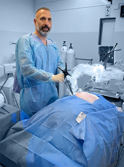
1. How is artificial intelligence transforming traditional surgical X-ray systems, and what specific capabilities distinguish AI-powered imaging from conventional ones?
To fully appreciate the impact of artificial intelligence, it is important to distinguish between two types of medical radiation: one used for diagnostic purposes and the other for treatment.
In the operating room, X-ray imaging is utilised to monitor the surgical process in real time. For example, when realigning a bone, X-rays are employed to ensure the bone is positioned correctly.
2. Can you explain how real-time AI image analysis supports surgeons during complex or time-critical procedures?
AI-powered image analysis can significantly enhance surgical decision-making. For example, when surgeons need to place a screw at a specific anatomical landmark—and the size or positioning of the screw may influence clinical outcomes—AI algorithms can provide precise, real-time evaluation and guidance. This enables optimal screw selection and placement, supporting the achievement of the desired surgical results.
3. What are the key AI algorithms or models most commonly applied in surgical X-ray interpretation, and how do they ensure diagnostic accuracy?
The key algorithm is yet to be determined due to the lack of experience and knowledge of the users.
Training approaches must be adapted to reflect the advanced technology implemented in the new CRM systems. Unlike legacy platforms, effective training should focus on evaluating measurements, trajectories, and precision, with content tailored to the specific requirements of each surgical procedure.
4. How does AI integration in X-ray systems help in detecting subtle anomalies that might be missed by the human eye?
Currently, we implement AI-powered tools for diagnostic imaging modalities such as CT, MRI, and other examinations. In the operating room, the primary objective is to evaluate the surgical process in real time and detect anomalies as they arise. To achieve this, it is essential to define the procedural workflow for the AI system. For example, in hybrid operating rooms used for angioplasty, AI can be configured to identify early signs of vascular rupture—enabling smart detection that can help prevent dangerous outcomes.
5. What measures are taken to ensure data integrity, security, and patient privacy when using AI-powered imaging systems in hospitals?
Measures implemented to ensure data integrity, security, and patient privacy include comprehensive data collection to validate the accuracy of information presented, secure connectivity through hospital firewalls and robust online security protocols, and the masking of patient data whenever system outputs are showcased or shared externally.
6. In what ways does AI help in reducing surgical risks or intraoperative complications compared to traditional imaging techniques?
Traditional imaging techniques rely heavily on the clinician’s interpretation and prior experience with similar cases. However, even when facing the same clinical problem, patient anatomy can vary significantly. In these situations, artificial intelligence can provide objective, real-time data to support the surgeon’s decision-making regarding the validity and course of the procedure.
It is important to emphasise that AI serves as an additional tool and cannot replace the surgeon. The ultimate responsibility for the success or failure of the operation remains with the surgeon, who must maintain accountability and view AI as a supportive resource rather than a substitute.
7. How can AI-enhanced imaging contribute to personalised surgical planning and precision-based decision-making?
That is an excellent question. In my experience, thorough surgical planning, understanding both the challenges and advantages of the procedure, is crucial for achieving optimal outcomes. Studies have shown that when surgeons are well-prepared and know what to expect, patient results tend to improve.
While the integration of AI into surgical planning is still evolving, these technologies will inevitably become an essential part of the process. By combining preoperative planning with real-time AI support during surgery, we can enhance intraoperative decision-making, reduce the duration of procedures, and ultimately improve patient outcomes.
For example, if a spine surgeon uses planning software to determine the optimal placement of screws, and the imaging AI can confirm in real time that the screws are positioned according to plan, this synergy can significantly contribute to the success of the procedure.
8. Could you elaborate on the workflow efficiencies gained through AI-driven automation in operating room imaging?
The workflow efficiencies achieved through AI-driven automation are similar to those provided by other supportive tools. In some cases, obtaining the optimal image requires the X-ray technician to repeat the image acquisition process multiple times, which increases exposure to X-rays for both the patient and the surgical staff. By integrating AI, the technician can make precise adjustments based on previously acquired images, thereby obtaining the best possible image for the surgeon while minimising unnecessary exposure.
For example, if there is a need to shift the image display to the left and it is difficult to do so manually, AI can be utilised to navigate the system to the desired location efficiently.
9. What are the current challenges in training AI models for surgical imaging, and how can these limitations be overcome?
In the traditional operating room imaging environment, training was often conducted informally, with experienced X-ray technicians mentoring newcomers. This approach was sufficient for legacy systems, where the technology was limited, and the primary requirement was simply to provide an image for the physician.
With the introduction of AI-powered imaging systems, however, training must become more formalised and comprehensive. Both X-ray technicians and surgeons need to be well-versed in the available tools and features to utilise them effectively during procedures. Additionally, the X-ray technician should be able to recommend the most appropriate features based on the specific needs of the surgeon.
10. How do regulatory and ethical considerations impact the deployment of AI-powered X-ray technologies in clinical settings?
For the purpose of regulating AI in surgical imaging, it is essential to strike the right balance. The use of AI during surgery is fundamentally different from its use in diagnostic X-rays. In the operating room, the surgeon must achieve the best possible outcome for the patient efficiently, and while AI can provide valuable support, it should not override the surgeon’s clinical judgment. Regulation of intraoperative AI imaging should consider that the surgeon receives additional feedback—such as the patient’s condition and real-time tactical information—beyond what the AI provides. AI should be regarded as a supportive tool rather than the primary objective of the procedure.
For example, if the AI indicates that a screw has been successfully inserted but the surgeon suspects otherwise, it is crucial that the surgeon relies on other available tools and clinical assessments to ensure the best outcome for the patient.
11. Can AI-powered X-ray systems adapt and improve through machine learning based on prior surgical data and outcomes?
When it comes to diagnostic imaging, there are virtually no limitations to the advantages that AI can provide, delivering faster results, more accurate interpretations, and ultimately better treatment for patients by leveraging data from various imaging modalities.
For treatment-oriented X-rays, the primary goal is to supply real-time data that assists the surgeon in making informed decisions about the surgical outcome. One area where this adaptive approach is particularly valuable is oncology: for instance, when a specimen is removed, and the surgical team needs to examine the margins, X-ray imaging of markers can be enhanced by AI to provide more accurate information about the specimen.
12. How does the collaboration between radiologists, surgeons, and AI systems evolve with the increasing integration of intelligent imaging tools?
From my experience, there are numerous ways in which collaboration between radiologists, surgeons, and other caregivers can be enhanced by AI. AI enables a seamless flow of patient results from one clinician to the next, involving all relevant caregivers almost immediately.
Whereas previously caregivers needed to be informed at each step, AI now delivers results the moment the patient completes the examination, ensuring that all parties are promptly updated and can coordinate care more efficiently.
13. What future innovations or research directions do you foresee for AI in intraoperative imaging and precision surgery?
Future innovation in this field is virtually limitless. Enhanced connectivity and improved accuracy will enable surgeons to deliver the best possible surgical care in minimal time, thereby reducing anesthesia duration, shortening operating room time, improving patient rehabilitation, and strengthening collaboration between departments.
In my view, surgeons will be able to share dilemmas with peers online and consult with AI regarding next steps if any uncertainties arise during surgery, based on the patient’s prior condition and medical history. For example, if there is a need to administer a specific drug, AI can provide recommendations on the most suitable option by considering patient information such as allergies or other relevant criteria.
14. In your opinion, how can hospitals ensure seamless adoption and clinician trust in AI-powered surgical imaging solutions?
In my opinion, experience and trust are fundamental. AI is intended to provide clinicians with greater confidence in their decisions, reducing reliance on guesswork. As these tools demonstrate reliability and accuracy, we can expect increasing dependence on AI-driven technologies, with certain procedures and workflows becoming based on their integration. Future surgeons are likely to rely on AI tools at every stage of a procedure. However, it is important to strike a balance between technological advancement and the development of clinical skills and knowledge.
We must also consider regulatory measures to ensure that caregivers possess the necessary skills and training to perform procedures independently, even as technology becomes more advanced. Precautions should be taken to use these technologies only in safe environments, allowing us to address potential setbacks effectively.