

Respiratory medicine encompasses acute disorders that require immediate assessment as well as chronic conditions that need regular monitoring. As a result, respiratory patients represent a substantial burden on healthcare systems worldwide. Traditional diagnostic pathways often rely on chest radiography and computed tomography (CT), which remain central thoracic imaging modalities but may be time-consuming, require patient transport, and expose patients to ionizing radiation.
Thoracic ultrasound offers a complementary approach that can be integrated with established imaging methods. Portable, repeatable, and radiation-free, it enables real-time bedside assessment as part of the clinical examination. Over recent decades, technological advances and growing clinical experience have transformed thoracic ultrasound from a limited diagnostic technique into an essential component of daily respiratory practice.
Recent international guidelines and consensus statements, including those from the European Respiratory Society, highlight the expanding role of lung ultrasound in respiratory medicine. Beyond diagnosis, ultrasound guides many interventional procedures within the thoracic cavity.
Why Thoracic Ultrasound Matters
Thoracic ultrasound is a safe, non-invasive tool that enables immediate, real-time patient assessment, unlike many conventional imaging techniques.
The technique is based on the interpretation of artifacts generated at the pleural surface, together with direct visualisation of structures such as pleural fluid, lung consolidations, diaphragmatic movement, and chest wall abnormalities. It can therefore identify clinically significant thoracic pathology while the patient remains at the bedside.
For healthcare organisations, the advantages of ultrasound extend beyond diagnosis. It supports clinical decision-making, improves procedural safety, and enables rapid bedside care in emergency departments, respiratory wards, intensive care units, and outpatient clinics.
Pneumothorax
Pneumothorax is one of the most important applications of thoracic ultrasound. Key sonographic findings include the absence of lung sliding, the barcode or stratosphere sign on M-mode, and identification of the lung point, which marks the transition between normally aerated lung and air within the pleural cavity.
Multiple studies have shown that thoracic ultrasound is more sensitive than chest radiography for detecting pneumothorax, particularly in emergency and critical care settings. This allows clinicians to make faster decisions after invasive procedures, trauma, or sudden respiratory deterioration.
In procedural settings, bedside ultrasound can reduce diagnostic delays and minimise risk while also providing a useful method for post-procedural assessment.
Pneumonia
Pneumonia can be effectively assessed with ultrasound, particularly when inflammatory changes reach the subpleural regions. Typical findings include subpleural consolidation, dynamic air bronchograms, B-lines, and tissue-like hepatisation of the lung.
Thoracic ultrasound is especially valuable when inflammatory changes extend to the pleural surface. In these situations, it can provide rapid bedside confirmation and help differentiate pneumonia from pleural effusion, atelectasis, and other peripheral lung abnormalities.
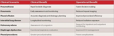
Beyond diagnosis, ultrasound can be repeated at the bedside to monitor disease progression and response to therapy.
Interstitial Syndromes and Interstitial Lung Disease
Thoracic ultrasound has become increasingly valuable in the assessment of diffuse parenchymal lung disease. The most characteristic sonographic finding is the presence of multiple vertical artifacts, commonly referred to as B-lines, which are caused by alterations in the subpleural interstitium.
B-lines typically indicate increased lung density, but they are not disease-specific and must always be interpreted in the clinical context. The differential diagnosis includes pulmonary edema, acute respiratory distress syndrome, and inflammatory conditions such as multilobar pneumonia.
In patients with interstitial lung disease, thoracic ultrasound may demonstrate diffuse B-lines, pleural-line irregularities, pleural thickening, and small subpleural consolidations. Several studies have reported correlations between ultrasound abnormalities and the extent of fibrosis identified on high-resolution CT.
One of the greatest advantages of thoracic ultrasound in interstitial lung disease is its potential for long-term monitoring. Serial examinations allow clinicians to assess disease progression, detect radiological deterioration earlier, and evaluate response to treatment.
Pleural Effusion
The evaluation of pleural effusion remains one of the most established applications of thoracic ultrasound and represents a cornerstone of modern pleural medicine.
Ultrasound is more sensitive than chest radiography for detecting pleural fluid. Importantly, it also allows assessment of fluid volume and characteristics, as well as adjacent lung abnormalities. A simple free-flowing effusion can be readily distinguished from a complex effusion with septations or pleural thickening.
Beyond diagnosis, ultrasound has fundamentally improved the safety profile of pleural procedures. Real-time image guidance allows precise site selection and reduces the risk of organ puncture and complications such as pneumothorax.
When integrated into routine pleural disease assessment, ultrasound improves diagnostic efficiency and procedural safety while reducing dependence on radiology resources.
Diaphragm Ultrasound
The diaphragm has traditionally been difficult to assess using conventional imaging techniques. Thoracic ultrasound now provides a practical, non-invasive method for evaluating diaphragmatic structure and function.
Clinicians can assess diaphragmatic position, excursion, thickening, and contractile activity. These measurements have become increasingly relevant in intensive care units, where diaphragm dysfunction is recognised as an important contributor to respiratory failure and prolonged mechanical ventilation.
Applications are also expanding in chronic respiratory diseases, neuromuscular disorders, and postoperative care, offering direct assessment of an important dimension of pulmonary function.
Ultrasound-Guided Procedures
Thoracic ultrasound has transformed the way pleural and pulmonary invasive procedures are performed.
Image guidance is now part of routine practice for thoracentesis, chest drain insertion, and pleural biopsy. By providing real-time visualisation of pleural fluid, lung tissue, and chest wall structures, ultrasound improves safety and reduces the risk of complications.
In addition to improving safety, ultrasound-guided procedures can support direct diagnosis and can be performed in a wide range of settings, including outpatient and ambulatory environments.
Implementation, Training, and Governance
The widespread adoption of thoracic ultrasound in respiratory medicine requires more than equipment acquisition. International guidance emphasises the need for structured training, competency assessment, and continuous quality assurance.
Healthcare organisations should establish governance frameworks that include image archiving, documentation standards, and regular competency review for clinicians. These measures help ensure consistency, reproducibility, and patient safety across clinical settings.
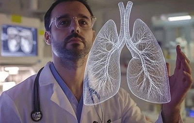
The increasing availability of handheld ultrasound devices creates further opportunities for broader implementation across emergency departments, respiratory units, intensive care services, and outpatient clinics.
Artificial Intelligence and Future Directions
Recent international consensus documents identify artificial intelligence as one of the most promising developments in thoracic ultrasound.
Emerging applications include automated B-line quantification, image-quality assessment, pattern recognition, and decision-support systems. These technologies have the potential to reduce operator variability, support less experienced users, and facilitate large-scale implementation.
Tele-ultrasound and online ultrasound platforms may further improve access by enabling remote interpretation and supervision. Future developments are also expected to focus on standardised reporting, quantitative ultrasound techniques, and integration with electronic health records.
Future Trends in Thoracic Ultrasound
• Handheld ultrasound devices integrated into routine clinical workflows and outpatient monitoring.
• Artificial intelligence-assisted image interpretation.
• Tele-ultrasound and remote patient monitoring.
• Integration with digital health platforms and electronic medical records.
Conclusions
Thoracic ultrasound is a safe, repeatable bedside diagnostic tool that has evolved into an essential part of modern respiratory medicine.
From the assessment of a wide range of pulmonary and pleural conditions to the guidance of complex procedures, thoracic ultrasound supports rapid clinical decision-making while improving diagnostic accuracy and operational efficiency.
As training pathways become more structured and technological advances such as artificial intelligence and tele-ultrasound continue to evolve, the role of thoracic ultrasound is likely to expand in daily clinical practice. Rather than replacing established imaging techniques, it should be used in combination with clinical assessment and other imaging modalities, providing immediate bedside information that supports high-quality patient care.
For healthcare organisations seeking to improve diagnostic efficiency, safety, and monitoring, thoracic ultrasound is one of the most effective technologies currently available.
References
1. Laursen CB, Clive A, Hallifax R, Pietersen PI, Asciak R, Davidsen JR, etal. European Respiratory Society statement on thoracic ultrasound. EurRespir J. 2021;57(3):2001519.
2. Demi L, Wolfram F, Klersy C, De Silvestri A, Ferretti VV, Muller M, etal. New international guidelines and consensus on the use of lungultrasound. J Ultrasound Med. 2023;42(2):309–344.
3. Volpicelli G, Elbarbary M, Blaivas M, Lichtenstein DA, Mathis G, Kirkpatrick AW, et al. International evidence-based recommendations for point-of-care lung ultrasound. Intensive Care Med. 2012;38(4):577–591.
4. Lichtenstein DA, Mezière GA. Relevance of lung ultrasound in the diagnosis of acute respiratory failure: The BLUE protocol. Chest. 2008;134(1):117–125.
5. Lichtenstein DA. Lung ultrasound in the critically ill. Ann Intensive Care. 2014;4:1.
6. Volpicelli G, Lamorte A, Villén T. What’s new in lung ultrasound during the COVID-19 pandemic. Intensive Care Med. 2020;46(7):1445–1448.
7. Mojoli F, Bouhemad B, Mongodi S, Lichtenstein D. Lung ultrasound for critically ill patients. Am J Respir Crit Care Med. 2019;199(6):701–714.
8. Mathis G. Thoraxsonography—Part II: Peripheral pulmonary consolidation. Ultrasound Med Biol. 1997;23(8):1141–1153.
9. Reissig A, Copetti R, Mathis G, Mempel C, Schuler A, Zechner P, et al. Lung ultrasound in the diagnosis and follow-up of community-acquired pneumonia. Eur J Radiol. 2012;81(5):e694–e700.
10. Long L, Zhao HT, Zhang ZY, Wang GY, Zhao HL. Lung ultrasound for the diagnosis of pneumonia in adults: A meta-analysis. Medicine (Baltimore). 2017;96(3):e5713.
11. Alrajab S, Youssef AM, Akkus NI, Caldito G. Pleural ultrasonography versus chest radiography for the diagnosis of pneumothorax: Review and meta-analysis. Crit Care. 2013;17(5):R208.
12. Eibenberger KL, Dock WI, Ammann ME, Dorffner R, Hörmann MF, Grabenwöger F. Quantification of pleural effusions: Sonography versus radiography. Radiology. 1994;191(3):681–684.
13. Lichtenstein D, Hulot JS, Rabiller A, Tostivint I, Mezière G. Feasibility and safety of ultrasound-aided thoracentesis in mechanically ventilated patients. Intensive Care Med. 1999;25(9):955–958.
14. Volpicelli G, Gargani L. Sonographic signs and patterns of COVID-19 pneumonia. Ultrasound J. 2020;12(1):22.
15. Gargani L, Volpicelli G. How I do it: Lung ultrasound. Cardiovasc Ultrasound. 2014;12:25.
16. Dietrich CF, Mathis G, Blaivas M, Volpicelli G, Seibel A, Wastl D, etal. Lung B-line artefacts and their use. J Thorac Dis. 2016;8(6):1356–1365.
17. Chiumello D, Mongodi S, Algieri I, Vergani GL, Orlando A, Via G, etal. Assessment of lung aeration and recruitment by lung ultrasound in acute respiratory distress syndrome patients. Crit Care Med. 2018;46(11):1761–1768.
18. Goligher EC, Dres M, Fan E, Rubenfeld GD, Scales DC, Herridge MS, etal. Mechanical ventilation–induced diaphragm atrophy strongly impacts clinical outcomes. Am J Respir Crit Care Med. 2018;197(2):204–213.
19. Via G, Storti E, Gulati G, Neri L, Mojoli F, Braschi A. Lung ultrasound in the ICU: From diagnostic instrument to respiratory monitoring tool. Minerva Anestesiol. 2012;78(11):1282–1296.
20. Rahman NM, Singanayagam A, Davies HE, Wrightson JM, Mishra EK, Lee YC, et al. Diagnostic accuracy, safety and utilisation of respiratory ultrasound in pleural disease. Thorax. 2010;65(5):449–453.