This topic is crucial for maintaining a safe environment for both patients, Companions and healthcare professionals in the field of radiology and imaging. Radiation Safety, MRI Safety, Contrast Safety, Infection Control, Safety during Interventional procedures.
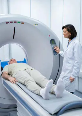
Radiology and imaging play a crucial role in modern healthcare, enabling doctors to make correct diagnoses and treat a wide range of medical conditions. However, like any medical procedure, radiology and imaging too involve risks, particularly related to radiation exposure, for both patients and radiologists or radiographers and healthcare workers.
It is important to reduce radiation-associated risk, as ionizing radiation used in various procedures like X-rays can deposit energy in human cells and cause tissue changes, increasing the risk of cancer, genetic mutations, and radiation-induced injuries like radiation sickness or acute radiation syndrome. Radiation sickness can cause symptoms like nausea and radiation burns. Hence, it is important to be conscious not only of the clinical advantages of medical imaging but also of the potential hazards and the safety measures that need to be rigorously adhered to. Ionizing radiation should not be used unless the scope of benefits it offers exceeds the risk to those who will be exposed to the radiation.
The risk posed by ionizing radiation can be minimised by applying the key principles in radiation protection. The focus should be on reducing or optimizing radiation exposure and customising it to address the individual study and patient requirements while also not compromising on imaging quality for accurate diagnosis.
The guiding principle of radiation safety is "ALARA" which stands for "as low as reasonably achievable." If there is no direct benefit from receiving a dose, even a small one, it should be avoided.
Time: Spend only as much time as needed near a radiation source. Stay only until the task is accomplished. Radiologists can use alarming dosimeters to help them minimise the amount of time they stay in an area with high radiation levels.
Distance: Increase the distance between the patient and radioactive source. That can help reduce the dose as well. But increasing the distance between the patient and the image receptor also increases the amount of scatter produced and fall in the image receptor, thus lowering image quality. The fluoroscopic exposure time has to be kept as short as possible, with focus on getting adequate images with the least exposure to the patient.
One also needs to ensure that repeated radiation exposure is not required. However, if need arises, the beam entrance port should be changed by rotating the tube around the patient, so that the same body surface area doesn't get exposed.
Shielding: Shielding involves the use of protective barriers between the radiation source and the operator, which may be structurally fixed, movable, or worn by operators. The best shielding is that which protects the whole body of an individual: walls, windows, doors, control booths, and x-ray tube housing. Protective clothing can shield operators from alpha and beta particles but will not protect them from gamma rays. Standing behind a wall can also serve as a shield, especially in cases of radiology emergencies.
Fluoroscopic tables with optional shielding and leaded drapes can help protect personnel from patient scatter. Protective clothing like aprons, vests, skirts, thyroid shields and gloves should be used while working in an unshielded environment. All these clothing items should be checked periodically for shielding integrity. However, shielding only protects from the effects of scatter radiation and not the primary beam. Radiation exposure to staff should be monitored using personnel dosimetry devices. The most commonly used ones are TLD badges. Badges should be worn at all times warranting radiation exposure or stored in a radiation free place when not in use. They should be worn inside the lead apron and not be shared with other individuals.
Leaded eyewear, like lead glasses or radiation glasses or opaque safety goggles can protect a worker's eyes from radiation exposure.
Radiation from medical examinations is similar to sunlight. The effect of sunlight on the skin depends on the light's intensity and how long a person stays in it. Hence, intensity, length of exposure and sensitivity of the skin need to be considered. According to the American College of Radiology, the approximate effective radiation dose for a CT scan is 7.7 mSv, which is equivalent to 2.6 years of radiation exposure in the natural background.
The number and length of digital acquisition or cine "runs" is one of the major components of patient radiation dose in interventional procedures. According to ALARA, the dose rate during cine runs is nearly 10 times the normal fluoroscopy dose rate. Cine runs should be avoided whenever possible, and the last image hold should be used instead for documentation.
Infection prevention and control (IPC) practices are important to prevent and control the spread of pathogens in medical imaging departments. Imaging rooms are used for both inpatients and outpatients, which often leads to contamination of surfaces, apparatuses and equipment. In MRI, the machine bore is the most common source of infection as it is difficult to access and usually overlooked during routine decontamination.
The waiting areas for patients and their family members can raise an inevitable possibility of infection transmission, particularly during outbreaks of highly contagious diseases like Covid-19.
According to the International Society for Infectious Diseases, here are some measures that should be taken to prevent or control infection spread.
• Strict adherence to hand hygiene
• Clean X-ray equipment and all other equipment with alcohol wipes or chlorhexidine-based disinfectant after every examination.
• Cover surfaces coming into direct contact with patients with a disposable sheet, which is changed after every scan.
• Wipes and alcohol gel (70% alcohol) should be used for the decontamination of radiographic markers, providing specific attention for ribbon markers.
• Disinfect the MRI machine with 500–2000 mg/L chlorine-containing disinfectant. Also, 2% double-chain quaternary ammonium salt or 75% ethanol can be used twice or more daily if it is not resistant to corrosion.
• Use the syringe, tube, and connector of the automatic injectors for only one patient.
• Regular surprise inspection of the hygiene of the imaging department is recommended so that it is ensured all hygiene practices are followed.
• Ensure an adequate supply of PPE.
• The surface of the CT equipment should be immediately decontaminated after the patient’s examination with solutions containing 5000-10,000 ppm available chlorine.42
• Emphasize environmental ventilation of examination rooms and if possible, disinfect air with hydrogen peroxide.
MRI has become an invaluable tool in the healthcare sector for vital diagnostic and anatomical information. While it avoids the use of ionizing radiation, it employs a strong magnetic field. Hence, MRI also has a unique set of risks and safety hazards that is very crucial to be aware of to avoid any untoward incidents. There are standardized guidelines for MRI site design, patient safety, and personnel workflow that need to be strictly adhered to ensure the safety of the patient and personnel.
Patients undergoing MRI are exposed to three different types of electromagnetic fields - static magnetic field, gradient magnetic field and radio-frequency electromagnetic wave, each with its own possible adverse effects. For instance, strong magnetic fields used in an MRI scan can cause heating of tissues, potentially leading to burns. Some patients may also experience anxiety or claustrophobia due to the enclosed environment and loud tapping, knocking or other sounds of the MRI machine.
Prior to an MRI scan, patients should be screened for safety risks such as implantable devices (pacemakers, ICDs) or other metals that may cause interference or health hazards. Patients should also change to radiation safe clothing before entering the radiation zone.
The MR imaging environment is divided into four distinct zones with progressive restriction of entry of people to avoid imaging related hazards. The restricted zones (zone III and IV) should always be under the direct supervision of people trained in MR safety. Any person who is not trained in MR safety protocols or not screened by MR personnel should be restricted from entering those zones.
All MRI facilities should have a documented plan to handle emergencies within zone IV, including cardiac arrest or code and fires, and all MRI personnel should be familiar with it. All the resuscitation and emergency equipment, including the crash cart or fire extinguishers should be verified as MR safe or MR conditional and should be placed in close proximity. During the event of an emergency, every effort must be made to move the patient and personnel outside zone IV, while preliminary resuscitation is begun by appropriately trained personnel.

During imaging tests, contrast agents (a group of chemical agents) may be used to distinguish one tissue or structure from its surroundings or to provide greater detail, improving diagnostic value of those imaging exams. Contrast agents from air to Barium to Iodine play a crucial role in X-ray and CT imaging. This inherent contrast allows radiologists and medical professionals to visualize the internal structures of the body and identify any abnormalities or pathology.
However, these drugs are also proven to cause allergic reactions and organ failure in susceptible patients. Therefore, it is important to know and understand safe use of contrast media.
All patients should be screened for risk factors for allergic reactions and renal dysfunction and explained to the patient, while obtaining consent. If any risk is involved, the risk-benefit profile of the proposed study should be considered for the patient prior to the administration of contrast material. If the risk of not performing the procedure can prove detrimental to the patient then the perceived risk from administering of contrast agent, the study should be carried out with following precautionary measures:
• Use the lowest possible dose of a low osmolar (Omnipaque) or iso osmolar (Visipaque) agent, for patients with cardiac disease with reduced ejection fraction.
• Inform the emergency department to have drugs and equipment on standby and be ready to treat any adverse reaction.
• Maintain close medical supervision during the entire study.
• Keep the patient under observation for 30 minutes after the procedure
• In case of patients with renal impairment, ensure they are well hydrated.
• In patients with risk factors, premedication with steroids can be tried to reduce the risk of reaction to contrast media. However, this method has not been proven to prevent acute allergic-like reactions.
• It is important to distinguish physiological reactions like nausea, vomiting, transient dizziness, hypertension, isolated chest pain, vasovagal reactions, arrhythmia, and convulsions from allergic reactions, as they don't require pre-medications for administering contrast media.
• All imaging centres where contrast material is used should have trained personnel who are equipped with the emergency supplies to treat any form of reaction.
• Contrast agents should be used with caution in the pediatric population as they have immature kidneys and lower GFR.
High-quality images help in making accurate diagnosis, planning the treatment and for medical follow-ups, thus helping in the patient's safety. One can obtain these high-quality digital images with a lower radiation dose by adjusting kVp, decreasing mAs, and decreasing focal spot size. Though a higher radiation dose leads to less noise and better image quality, one should be very cautious about the radiation dose. Hence, the radiographer should be well aware of the safety guidelines and use the radiographic systems in an optimized manner to obtain image quality that offers accurate diagnosis, but at the least possible radiation dose.
Ensuring safety in radiology and imaging is a shared responsibility between patients, radiologists, healthcare professionals and imaging facilities. By understanding the risks associated with radiation exposure and following best practices for safety, the radiation associated risks can be minimised and it can be ensured that radiological procedures are performed more effectively.
References:
• L.A.R.A informative pages magazine (November 2019 edition)
• Radiation dose to adults from common imaging examinations, American College of Radiology
• The International Society for Infectious Diseases - Guide to Infection control in the healthcare setting; Infection prevention and control in radiology department/ services (https://isid.org/guide/hospital/infection-prevention-and-control-in-the-radiology-department-service/)
• Radiographic Contrast Agents and Contrast Reactions (https://www.msdmanuals.com/professional/special-subjects/principles-of-radiologic-imaging/radiographic-contrast-agents-and-contrast-reactions)
• Tompe A, Sargar K. X-Ray Image Quality Assurance. [Updated 2022 Oct 17]. In: StatPearls [Internet]. Treasure Island (FL): StatPearls Publishing; 2024 Jan-. Available from: https://www.ncbi.nlm.nih.gov/books/NBK564362/

Anto Ramesh Delvi D is the Chairman and Managing Director of RADBLOX Healthcare Services Pvt. Ltd., with nearly three decades of experience in hospital care, home healthcare, radiology, and teleradiology across countries such as Indonesia, various parts of Africa, the Maldives, and Mauritius. Skilled in AI, medical laws, and hospital administration, he stays up-to-date with the latest technologies in the healthcare industry. Beginning from humble origins, he led Columbia Asia Hospitals Radiology Group, expanding it to 12 hospitals in eight cities across three countries. Anto is also a trusted health education trainer and industry expert.